Ocular trauma involves injuries to the eye and surrounding structures. In children, this presents unique challenges in evaluation, management and long-term outcomes. Experts from various parts of the world contribute their expertise in preserving visual function and addressing potential complications on Day 2 of the 5th World Congress of Paediatric Ophthalmology & Strabismus (WCPOS V 2024).
Risks and prevention
First on the podium was Dr. Inez Wong (Singapore), who presented on the epidemiology and mechanisms of pediatric ocular trauma. She noted that every year, 160,000 to 280,000 of children below 15 years old were hospitalized for ocular trauma. At the severe end of the spectrum, 21-24% involved penetrating globe injuries.1
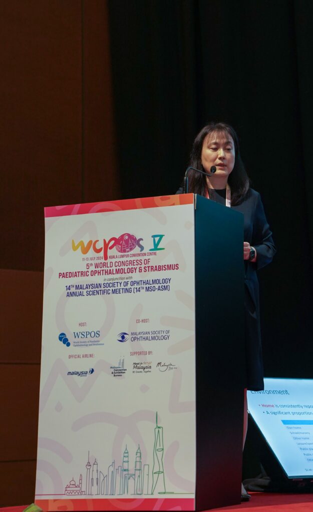
“There is a bimodal distribution among preschool children and teenagers, and males are at the highest risk of injury,” said Dr. Wong. This is attributed to the development of fine and gross motor skills in younger children and participation in sports or risky behaviors among older adolescents.
She highlighted that more injuries occur at home than in school and the outdoors, with closed globe injuries predominating the cases. Additionally, there is a high risk of refractive error, amblyopia and a poor visual prognosis associated with penetrating eye injuries in young children.
In conclusion, she offered suggestions for prevention, including adult supervision during play times, especially with toys that have projectile components, educating parents on hazards in the home, mandating the use of sports protective eyewear, and enforcing restrictions on airguns and fireworks.
Use of stent in canaliculi repair
Presenting on pediatric eyelid trauma, Dr. Jamalia Rahmat (Malaysia) emphasized that the goal of trauma reconstruction is to restore normal function as much as possible. Knowledge of the anatomic path of canaliculus is crucial and magnification with the microscope or surgical loupe would help in the identification of the medial cut end of the lacerated canaliculus.
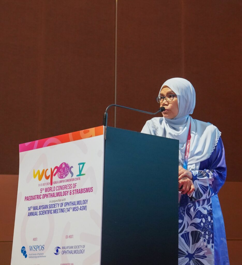
“The use of stent in canaliculi repair helps to keep the tissue together and makes your suturing much easier. It will also facilitate mucosal healing of the anastomosed canalicular and prevent canalicular obstruction/stenosis,” she said.
She stressed that all canalicular cuts should be repaired, noting that delayed repair (up to 48-72 hours) is not necessarily a bad thing, if that would allow for a more experienced surgeon, a more controlled situation, less edema and better visualization. Monocanalicular stent is easier to handle than the bicanalicular stent.
As for suturing the canicular tissue, Dr. Rahmat said: “Direct suturing that is not done properly may cause problems such as more scarring and stenosis. So, all you need is probably just to purse the string around the canicular to hold the stent there, which would be good enough. Maintain the stent in place for at least 6 weeks. Then, assess the functional success postoperatively.”
Repairing globe rupture
Speaking on globe rupture, Dr. Naomi Tan (United Kingdom) stressed the importance of taking an accurate history from the child or witness to establish the likelihood of an open globe injury. High-risk mechanisms include high-force blunt injury, high-velocity projectiles, sharp objects, foreign bodies and contamination, she noted.
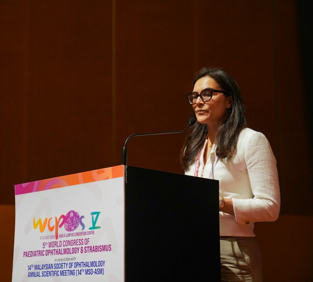
Dr. Tan explained that while assessing presenting visual acuity (VA) is important, it should not overly influence management decisions in children. Studies have shown no difference in presenting acuity between closed and open globe injuries in children. Additionally, adult studies have indicated that patients with no perception of light can recover.
She emphasized that surgical principles to follow when repairing a globe rupture in children include applying minimal pressure on the globe, providing maximum exposure, opposing identifiable edges and preserving tissue where viable, which can be achieved with sutures, glue or tectonic grafts.
“Be extremely careful when examining awake patients. Have a low threshold for examination under anesthesia and primary repair, and beware of occult injury,” she cautioned.
Strategies in managing traumatic cataracts
Next, Prof. Jaspreet Sukhija (India) offered tips to address traumatic cataracts. “Traumatic injury to the lens can involve other ocular structures, increasing the complexity of surgery. Increased incidence of inflammation and other sequelae can also adversely affect visual outcomes in the amblyogenic age group. That’s why it’s important that we tackle the cataract early,” he stressed.
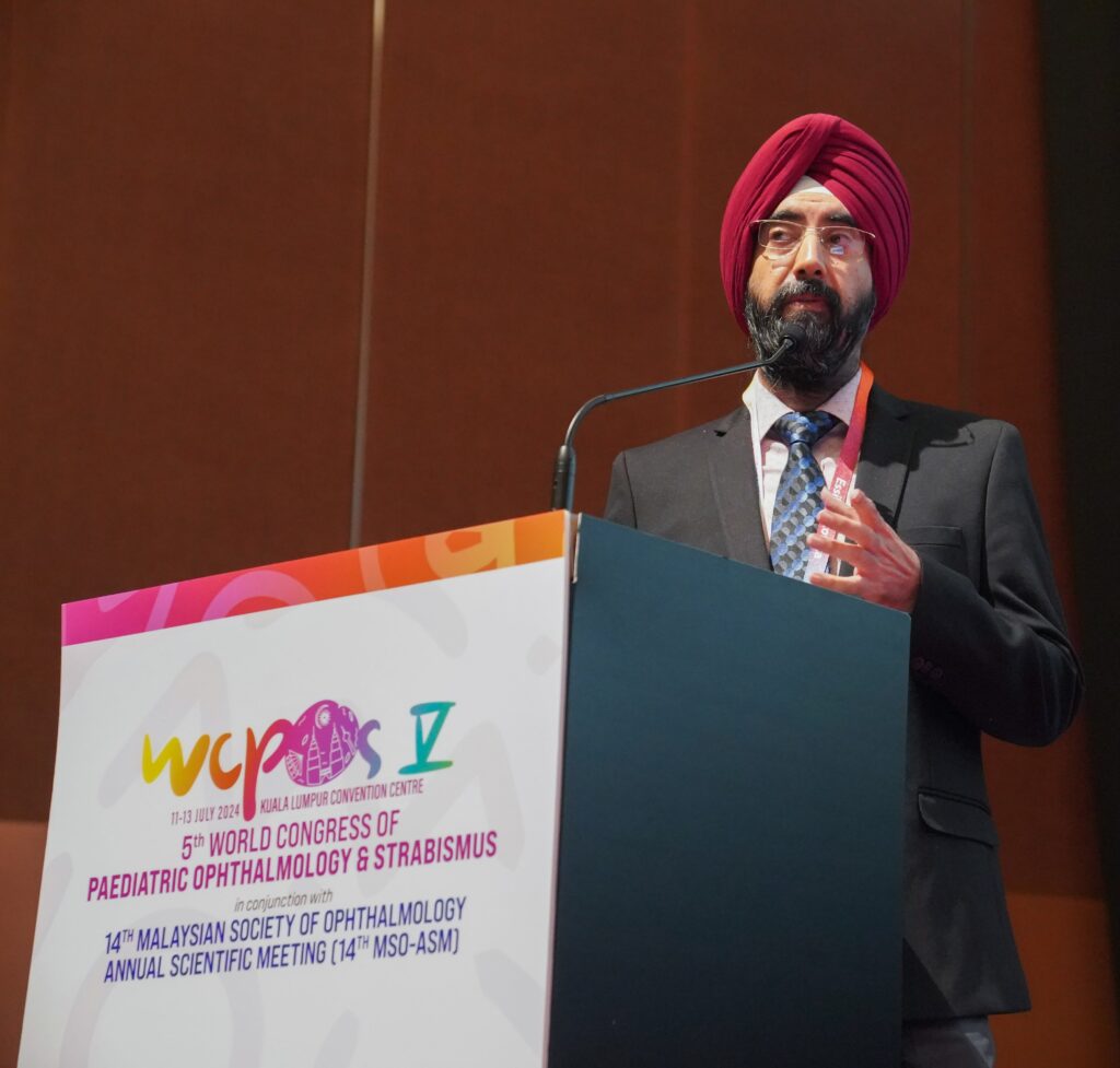
He noted that accurate intraocular lens (IOL) power calculations are challenging, and the debate continues between primary and secondary IOL implantation.
To obtain optimal outcome in such cases, he emphasized that preoperative ultrasound (USG) is crucial for assessing the posterior segment and the posterior capsule’s condition. Biometry should be performed on the same eye whenever possible. Meanwhile, the staging of primary IOL implantation depends on whether the injury involves an open or closed globe.
“Always stain the anterior capsule for better management. And be wary of the vitreous and the shaky bag,” he added.
Furthermore, Prof. Sukhija advised on performing a posterior primary capsulorhexis (PPC) due to the high incidence of visual axis opacification (VAO) in children. Additionally, he suggested that posterior optic capture can prevent iris capture and synechiae formation, and intracameral steroids can help to reduce inflammation.
Management of strabismus following trauma
According to Dr. Rasheena Bansal (India), strabismus can develop in children following orbital or ocular trauma, although such cases are less common compared to other causes. This condition can manifest as both restrictive and paralytic in nature. Approximately, 5-10% of children who experience significant trauma develop strabismus.2
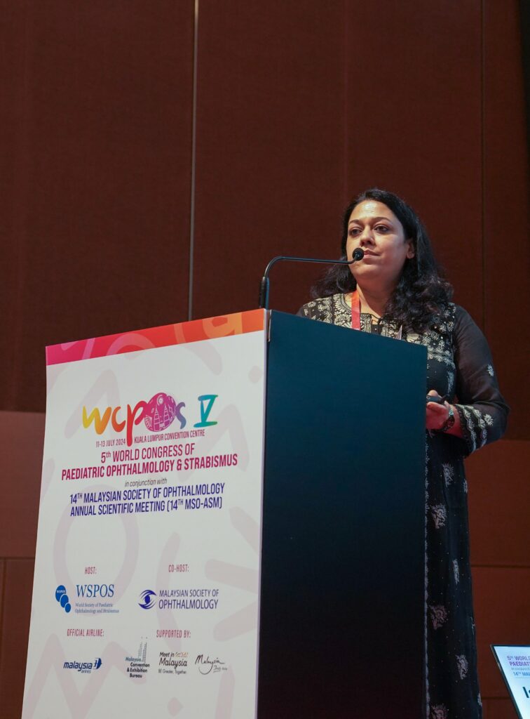
She explained that trauma can lead to strabismus due to direct leash effect (caused by accessory scar tissue between globe and orbital wall, or tight extraocular muscle) or reverse leash effect (where the muscle is fixed to the globe due to scarring or with suture), restricting muscle mobility.
Regarding treatment, Dr. Bansal recommended initially opting for a conservative approach involving non-surgical methods such as patching, prisms and vision therapy. “Surgical intervention is indicated when misalignment persists despite non-surgical efforts, when there is significant deviation affecting vision and quality of life, when double vision persists despite treatment, or when functional amblyopia develops due to vision loss in one eye. Surgical options include recession/resection, transposition and adjustable suture techniques,” she said.
In conclusion, Dr. Bansal emphasized the importance of early diagnosis and treatment. “It is important to intervene early depending on the severity of the trauma, so that we can maximize the visual outcome and prognosis. Every case needs a different approach based on clinical presentation,” she said.
References
- Abbott J, Shah P. The epidemiology and etiology of pediatric ocular trauma. Surv Ophthalmol. 2013;58(5):476-485.
- Bhate M, Adewara B, Bothra N. Strabismus in pediatric orbital wall fractures. Indian J Ophthalmol. 2023;71(3):973-976.
Editor’s Note: Reporting for this article occurred at the 5th World Congress of Paediatric Ophthalmology & Strabismus (WCPOS V 2024) from 11-13 July in Kuala Lumpur, Malaysia.


![iStock 1207202567 [Converted]_V2](https://cookiemagazine.org/wp-content/uploads/sites/6/2022/03/iStock-1207202567-Converted_V2-01-579x326.jpg)
